Vascular Clog Loss Classification: An Advanced Alzheimer’s Research Using ConvNets
In this article, I will be discussing the paper I worked along with my peers Ambarish Moharil, Chirag Kedia, and Nikhil Sonavane. In the research paper we have discussed new algorithms and frameworks that could be used to classify an Alzheimer's brain from a normal healthy brain with an accuracy of 98.5 %.

Alzheimer’s disease is a neurodegenerative, irreversible, chronic brain disease that starts slowly and progressively worsens over time, destroying the brain cells leading to loss of cognitive and thinking skills which then ultimately lead to a state where patient can’t even perform simple diurnal tasks
Early diagnosis of AD would facilitate an early treatment intervention and family planning, as the studies have found out that if early diagnosis and treatment of AD would ultimately lead to saving $7.9 trillion dollars annually. But the main issue here is that diagnosis of AD is a very intense, vigorous, time-consuming, arduous task and significantly a very costly process involving a lot of mental and physical exams, followed by some laboratory and neurology tests, along with some neurological imaging (using MRI or CT scans). The cost of a brain MRI could very easily range from $500 to $1500. This cost is very unfeasible for families that are uninsured or underinsured.
Some of the techniques being previously employed for Alzheimer detection are Neural Networks (NN), KNN, and Support Vector Machines (SVM). Some of the deep learning methods can also be used such as Convolution Neural Networks (CNN), Long Short-Term Memory (LSTM), Recurrent Neural Networks (RNN), Generative Adversarial Networks (GANs) and Extreme learning, etc.
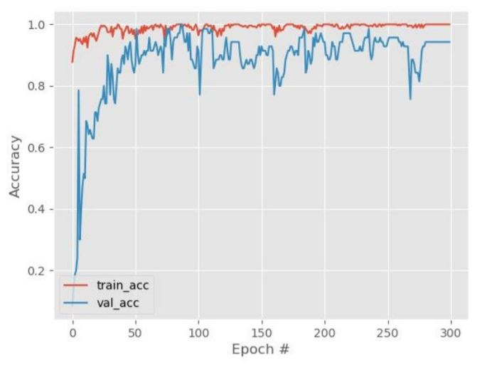
Flow Diagram

The steps that we have followed from collection of data, data processing and cleaning and our analysis.
- The dataset consisted of 4600 videos of cranial vessels classified as flowing or stalled
- After extracting the frames and manual filtering of the images, a dataset of 78675 images in total was created. If we observe the dataset in a scrupulous manner, we find out that the difference between the images as a whole is quite inconsequential. The difference between both the classes viz. blood flowing and stalled in the cranial vessel can be determined only by the region outlined in these images. So, unless and until we consider just the outlined region, the difference in both the classes seems irrelevant. Hence, extraction of the outlined region in a particular image becomes extremely important.

3. Furthermore, once the frames were extracted from the videos, these images were cropped to train the classifier. Regions of blood flow and stalled vessels were randomly cropped from the extracted frames.

4. This exercise was performed to train the classifier in a more efficient way by omitting the unnecessary parts and extracting the necessary data from the images.
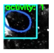
5. The above images where then classified using four distinct architectures viz. Resnet50, Resnet101, Resnet152 and AlexNet. The logic that is used in the classification is that in case when blood is actually flowing through the vessel a luminescent spot is picked up by the algorithm whereas in case when the blood is not flowing or stalled a black spot can be observed. So when we are looking at images of a non Alzheimer individual the frequency of these bright luminescent spots would be higher as compared to the black spots and vice versa for an Alzheimer infected person. Whichever algorithm is able to best pick up this difference will give us the highest accuracy
Result
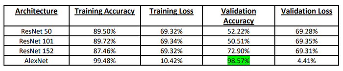
AlexNet was seen to give the best performance with a huge accuracy of 98.57%. Not only the accuracy is high, but the losses are low as well. The AlexNet model helped hugely in predicting accurately a person affected by Alzheimer’s.
